Imaging Centre
Services and equipment supporting electron microscopy, atomic force microscopy, light microscopy, histology, x-ray crystallography.
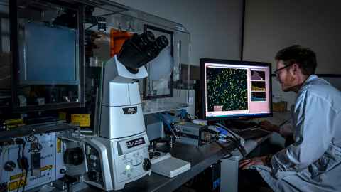
Equipment list
Transmission Electron microscopes
- FEI Tecnai FEG20 (200 kV, cryo-equipped, with STEM, EDS, EELS/EFTEM)
- FEI Tecnai 12 (120 kV, cryo-equipped)
- Philips CM12
- Sample preparation equipment
Atomic Force microscopes
- Asylum Cypher ES
- Asylum MFP-3D Origin
Light microscopes
- Andor Revolution spinning disc confocal/TIRF/ live cell microscope
- Leica LMD6000 laser microdissection microscope
- Nikon Ni-E motorised fluorescence microscope
- Nikon Ti-E motorised inverted fluorescence microscope
- Leica DMR upright fluorescence microscopes (2)
- Nikon Ni-U upright fluorescence microscope
- Leica Macroscope fluorescence zoom microscope
- Leica M80 stereo microscope
- Image processing software
High-content imaging system
- PerkinElmer Operetta
Histology equipment
- Leica CM1850 cryostat
- Tissue-Tek VIP 2000 automatic wax infiltration processor
Slide scanner
- PerkinElmer Vectra Polaris
X-ray diffraction
- Rigaku XtaLAB Synergy-S
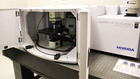
Horiba Labram HR Evolution Confocal Raman Spectroscope
Transmission Electron microscopes
The Centre specialises in transmission electron microscopy, and has close links with the research group of Associate Professor Alok Mitra. The electron microscopy equipment includes a broad range of preparation facilities for conventional and frozen samples, in addition to the microscopes themselves.
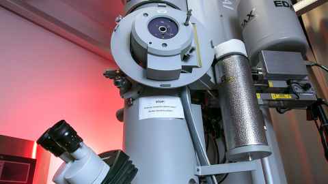
FEI Tecnai FEG20
The Tecnai FEG20 TEM is operated jointly with the Faculty of Engineering. This 200 kV TEM is equipped with a field emission electron source, STEM imaging mode, Gatan energy filter and Edax EDS. Gatan Ultrascan 1000 4-Mpixel digital cameras record conventional and energy filtered images, and a Fischione high-angle annular dark field detector (HAADF) records dark field images. Sample holders include conventional and double-tilt room temperature, and standard and high-tilt cryo holders. Tomography software is available.
Features of the Tecnai FEG20 include:
- High accelerating voltage (200 kV) for increased resolving power, greater penetration
- TEM or STEM mode
- Z-contrast imaging with HAADF
- EDS (X-ray spectroscopy) for elemental analysis
- Automated tomography in TEM or STEM mode
- Cryo-TEM with high-tilt stages and anti-contaminator
- Gatan energy filter (EELS) for elemental analysis, image filtering, fine probing of states of matter.
FEI Tecnai 12
The 120 kV Tecnai 12 TEM is optimised for cryo-electron microscopy, with Gatan cold stages and software that facilitates low dose imaging. It is equipped with a Gatan Ultrascan 1000 4-Mpixel digital camera and can also use plate film.
Features of the Tecnai 12 include:
- 120 kV for high contrast and resolution
- Cryo-TEM
- Bottom-mount digital camera for high resolution imaging
Philips CM12
The 120 kV CM12 TEM is ideal for routine scanning and imaging of biological and materials science specimens. It is equipped with a Gatan Bioscan 1-Mpixel digital camera.
Features of the Philips CM12 include:
- 120 kV for high contrast and resolution
- Top-mount digital camera for wide-field imaging
Sample preparation equipment for TEM
The Imaging Centre is equipped with a wide range of ancillary equipment for preparation of both biological and materials science TEM specimens, as well as for analysis of images, including:
- sample vitrifier (FEI Vitrobot) for cryo-TEM
- high-tilt cryo holders with pumping stations (Gatan)
- tomography holders (FEI and Fischione)
- double-tilt low background holder (FEI)
- glow discharge apparatus (EMS GloQube)
- plasma cleaner (Fischione)
- carbon evaporator unit (Edwards)
- ultramicrotome (Leica Ultracut)
- optical diffractometer
- ion mill (Fischione)
- dimple grinder (Fischione)
- ultrasonic disc cutter (Fischione)
Atomic Force microscopes
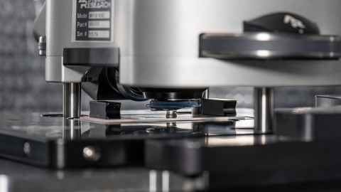
Atomic Force Microscopy is a very high-resolution type of scanning probe microscope with a resolution in the order of fractions of a nanometre. The sample is scanned by “touching” the surface with a mechanical probe and, by varying the type of probe used and the way that it interacts with the sample surface, a wide variety of information can be gained:
- Produce 3D models of your sample surface from the micrometre to nanometre scale
- Quantify roughness, depth profiles and grain sizes
- Map the mechanical properties of your sample: contrast materials with different elastic moduli and produce force-distance curves at selected spots
- Map the electrictronic properties of your sample: contrast materials with varying conductivity or map the piezoelectric response of your sample
- Map the magnetic properties of your sample: identify different magnetic domains on the surface of your sample
- In situ control of temperature and sample atmosphere composition.
There are many fields that can benefit from advanced AFM techniques including materials and polymer sciences, engineering, life sciences, thin film and functional surfaces research.
Cypher ES Environmental AFM
Cypher was the first commercial AFM to take advantage of the high bandwidth and low noise performance of small cantilevers and is the only commercial AFM to routinely resolve atomic point defects in the DNA helix. These capabilities were extended to a wider range of samples with the Cypher ES, which adds full environmental control to the Cypher platform.
Our Cypher ES has a scan range of 20*20 μm and is capable of the following AFM techniques:
- Imaging: Contact mode, Tapping/Non-contact mode, Frequency modulation, Phase imaging and Dual-AC mode*
- Nanomechanical: Force mapping, Force curves, AM-FM/Loss-tangent*, Force modulation and LFM
- Nanoelectrical: SKPM*, EFM* and PFM
- Magnetic force microscopy*
- Nanolithography and nanomanipulation.
*Requires specialised AFM probe.
Our Cypher ES also supports multiple sample environments:
- Measurements in air and in static liquid
- Gas perfusion in a sealed cell
- In-situ temperature control (0-120°C)
- Broad chemical compatibility
MFP-3D Origin
The MFP-3D Origin is capable of handling larger and rougher samples that the Cypher-ES while still being capable of high resolution imaging in both air and fluid environments.
Our MFP-3D Origin has a scan range of 90*90 μm and is capable of the following AFM techniques:
- Imaging: Contact mode, tapping/non-contact mode, frequency modulation, phase imaging
- Nanomechanical: Force mapping, force curves, force
modulation, LFM - Nanoelectrical: PFM
- Magnetic force microscopy*
- Nanolithography and nanomanipulation
*Requires specialised AFM probe.
Our MFP-3D Origin also supports multiple sample environments:
- Measurements in air and in static liquid
- In-situ electrochemical cell
- Micrometre control of the sample stage allows multiple adjacent scans to be combined into a larger surface model.
Light microscopes
A wide range of well-equipped light microscopes are available. Our microscopes range from basic zoom stereo microscopes through upright and inverted models equipped for epifluorescence, dark field, and multiple bright-field modes, to fully automatic models equipped for laser microdissection, confocal and live cell imaging. Each comes with colour or greyscale high quality digital cameras (or both). Bright field modes include phase contrast, DIC and modulation contrast. For fluorescence microscopy, a wide range of filter cubes is provided.
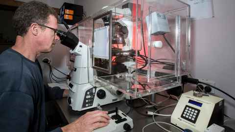
Andor Revolution spinning disc confocal microscope
This microscope is optimised for long-term and/or high speed imaging of, in particular, live cell specimens. The spinning disc confocal system allows faster imaging and lower overall illumination levels than is practical with conventional point scanning confocals.
Four laser lines (violet, blue, green and red) and a wide range of emission filters offer flexibility in design of experiments. A total internal reflection (TIRF) module offers enhanced resolution of cell surface features and isolated molecular assemblages.
An incubator cabinet allows for temperature regulation and control of CO2 supply to the specimen.
Cameras: Andor Ixon 885 (1-Mpixel EMCCD); up to 30 full frames per second; dual Andor Zyla (4-Mpixel sCMOS)
Software: Nikon Elements, Andor iQ
Leica LMD6000 Laser microdissection system
A powerful laser combined with a fully automated compound microscope allows the operator to speedily excise and collect tiny regions of interest from a slide-mounted specimen. The isolated fragments can then be analysed by PCR and mass spectroscopy. Fluorescence imaging is accommodated.
Nikon Ni-E motorised fluorescence microscope
Full motorisation and excellent optical components allow for efficient and sensitive imaging, especially in fluorescence mode. Image tiling is simple and reliable.
Cameras: Andor Zyla (5.5-Mpixel sCMOS).
Software: Nikon Elements
Nikon Ti-E motorised inverted fluorescence microscope
Semi-automatic (lenses, focus, filter cubes). Flexible imaging of plates, dishes and slides using fluorescence, phase, DIC and modulation contrast.
Cameras: Andor Clara (1.4-Mpixel CCD); Nikon DS-Ri-1 (up to 12 M-pixel colour CCD)
Software: Nikon Elements
Leica DMR upright fluorescence microscopes (2)
Two all-round upright microscopes fitted with new colour CMOS cameras and equipped for phase, DIC and fluorescence.
Camera: Progres Gryphax (2.3- and 5-Mpixel colour CMOS); Spot Pursuit (1.4-Mpixel CCD)
Nikon Ni-U upright fluorescence microscope
A specialist fluorescence tool equipped with sensitive lambda lenses and dual metal halide and xenon light sources.
Camera: Spot Pursuit (1.4-Mpixel CCD)
Leica Macroscope fluorescence zoom microscope
For lower magnification (6 – 90x) imaging, including fluorescence.
Camera: Leica DC-500 (1.4-Mpixel colour CCD)
Leica M80 stereo microscope
One of several stereo microscopes, this one has multiple illumination options including shadowless.
Camera: Lumenera Infinity (2-Mpixel colour CCD)
Image processing software
A variety of packages from ImageJ through Amira and Metamorph to Bitplane Imaris.
High-content imaging system
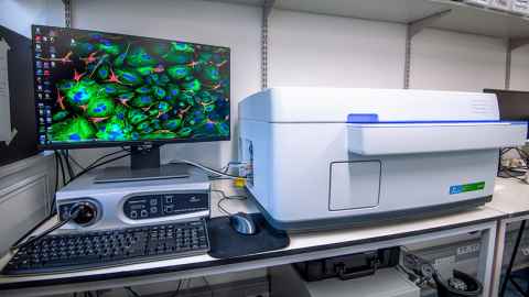
Fully automated image acquisition of live or fixed specimens using slides or multi-well plates.
PerkinElmer Operetta
The Operetta has three imaging modes – bright field, digital phase contrast, and fluorescence, with a confocal imaging option to eliminate background. Featuring a live cell chamber (37°C and CO2 control), enabling controlled time course imaging at 2, 10, 20 & 40X magnification.
Histology equipment
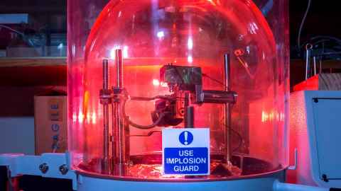
A full histology service is available for paraffin- and cryo-embedded specimens.
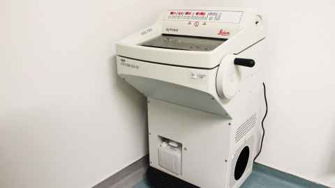
Leica CM1850 cryostat
For the preparation of frozen sections.
Tissue-Tek VIP 2000 automatic wax infiltration processor
Also available are a wax embedding station and rotary microtome for the preparation of paraffin sections. A dedicated staining run facilitates H&E processing.
A vibratome and a sledge microtome provide options for sectioning challenging specimens.
Slide scanner
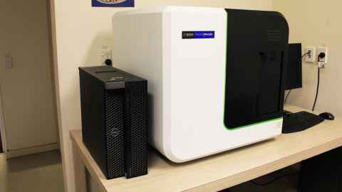
PerkinElmer Vectra Polaris
PerkinElmer (now Akoya Biosciences) Vectra Polaris slide scanner is now available in the imaging centre. The Vectra Polaris can be used for bright field slide scanning, multi-colour fluorescence slide scanning and multi-spectral slide scanning.
Using multi-spectral imaging the system can separate up to 9 different colours as well as separating autofluorescence background from the real signal. The system can also be used with filter cubes to separate up to 5 different colours. Vectra Polaris is optimised for Akoya Biosciences Opal assay kits but other fluorescent markers can be used as well.
Features of the Vectra Polaris include:
- Resolution: 10x (1.0 um/pixel), 20x (0.5 um/pixel), 40x (0.25 um/pixel)
- Detection method: Fluorescence, Brightfield
- Tissue formats: tissue sections and tissue microarrays
- 80 slide capacity with continuous loading technology
- Automation: touchless, with walk-away image acquisition
- Inform image analysis software: intuitive learn-by-example interface to automatically segment and quantitate tissue structures, cells and sub-cellular signatures
X-ray diffraction
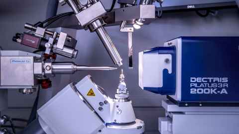
Collection of crystallographic data for structural characterisation of proteins.
Rigaku XtaLAB Synergy-S
The Rigaku XtaLAB Synergy-S X-ray diffractometer enables single crystal X-ray diffraction measurements. The system comprises a Microfocus X-ray source, a PILATUS4 R 200K single photon counting detector, and an Oxford Cryosystems Cryostream 800, allowing accurate control of the sample temperature. It is also equipped with a plate holder attachment for in situ diffraction of crystals in crystallisation plates.
How we can help you
Staff are on hand to provide initial training as well as ongoing support and advice as required.
The centre staff will carry out projects on behalf of staff and students when it is cost and time-efficient to do so. There may be charges for this service.
Where time permits, we are happy to train users fully in the operation of the unit’s equipment and facilities, and to make equipment available for independent use.
Booking and access
Our microscopes are available for booking through iLab.
Project design/method development
You are advised to consult with Centre staff in good time before deciding on your imaging strategy. Standard methods are available for all instruments and techniques, but it is commonly the case that minor or major modifications or innovations may be required to ensure the success of the imaging project.
For cost information and specific sample requirements please refer to the individual equipment.
Alternative or related places to go
Scanning electron microscopy: The Research Centre for Surface and Materials Science (RCSMS), Department of Chemical and Materials Engineering.
Computerised tomography (micro CT) X-ray scanning: Auckland Bioengineering Institute (ABI)
Further light and electron microscopy options: Biomedical Imaging Research Unit (BIRU), Faculty of Medical and Health Science
Our people
- Manager imaging centre | Dr Iain Hay
- Histologist | Jennifer Chen
- Manager light and electron microscopy | Dr Adrian Turner
- Slide scanner | Dr Saem Park
- Technical support for Raman spectroscopy | Dr Cherie Tollemache
- Technical support light microscopy | Dr Charlie Johnson
- Technical support atomic force microscopy | Joseph Vella
- Manager x-ray diffraction facility | Associate Professor Chris Squire
- Technical support for cryo-TEM and micro electron diffraction | Dr Paul Young
- Technical support for HRTEM and sample preparation of hard materials | Dr Shanghai Wei